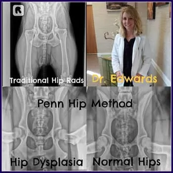
The most commonly inherited orthopedic disease, hip dysplasia afflicts many large breed dogs resulting from loose and poorly configured hip joints. As these dogs age, the hip joints more readily develop osteoarthritis that can cause pain and limit their mobility.
The Penn HIP screening is a set of three x-rays of the hips performed under heavy sedation or anesthesia that can assess dogs as young as 16 weeks old for signs of hip dysplasia. This process has been scientifically shown to be one of the best ways to assess dogs for hip dysplasia and has a great deal of interest from many breed organizations. The three x-ray views include a standard hip view to examine for osteoarthritis, a compressed view to assess hip joint quality of fit, and a distracted view to measure the degree of joint laxity (looseness). These x-rays are used by Antech radiologists to evaluate the quality of hip conformation and also assign a number (distraction index) to designate the degree of hip laxity. Dog breeders may use this evaluation to help select breeding stock to reduce the prevalence of the disease in their lines, and dog owners may use this screening to determine the need for early medical, surgical, or environmental management strategies of young dogs that may be affected. Early intervention with this disease can allow reduction in the clinical symptoms of hip dysplasia long term, helping our dogs live healthier and more comfortable lives.
To perform Penn HIP, a veterinarian must become certified, which requires coursework as well as technical training in the specific radiographic technique. Here at Patton Veterinary Hospital, Dr. Stephanie Edwards has completed her Penn HIP certification and would love to evaluate your pet. If interested in having this screening procedure done for your dog, please contact us for more information. If you would like more information on Penn HIP, please visit: http://info.antechimagingservices.com/pennhip/index.html .
