Glaucoma in Dogs and Cats
- posted: Sep. 23, 2023
Glaucoma in Pets
Glaucoma occurs when the pressure inside the eye increases which leads to pain and loss of vision. Glaucoma is a relatively rare disease and it is more common in dogs than in cats. The condition can be caused by a primary anatomic or chemical defect in how fluid drains from the eye i.e., a “clogged” sieve or it may be secondary to a medical condition such as a tumor or chronic inflammation. Secondary glaucoma is more common in cats. Primary glaucoma can occur as a hereditary condition in many breeds of dogs including the cocker spaniel, beagle, basset, Samoyed, Shih Tzu and Chinese Shar Pei. Siamese and Burmese cats may have an increased risk of primary glaucoma.
Normal eye pressure is a function of balance between production of normal eye fluid called aqueous humor and drainage of this fluid within the eye. If the sieve-like structure between the cornea and iris does not function properly, fluid cannot drain and eye pressure increases. Glaucoma is always caused by decreased drainage, not by increased fluid production.
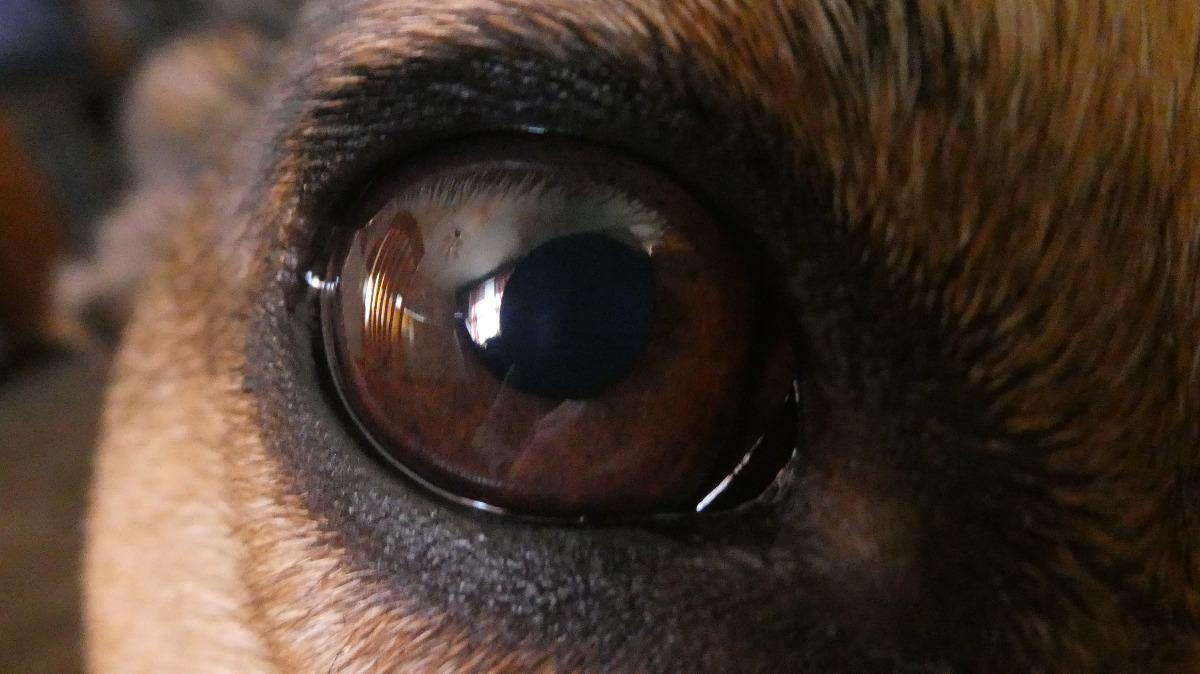
Early or mild glaucoma may not produce any noticeable physical changes; however, as the pressure increases, the eye will appear to grow larger or bulge and the pet may experience pain, shying away from touch around the head or face and exhibiting increased squinting or blinking. The eye may also look quite red and the pupil may be larger or dilate on the affected side. Pets may also bump into things as a result of lost vision in the affected eye. Glaucoma usually begins in just one eye, but, if due to a genetic or inherited condition, the other eye may also be affected in time.
If glaucoma is suspected or if your pet is a high-risk breed, the pressure inside the eye can be measured with a device called a tonometer. A drop of numbing medicine is applied to the cornea and the tonometer is touched repeatedly to the surface of the eye to obtain a reading. It looks a bit odd and some dogs and cats resist testing, but it is not painful and is relatively quick to perform. If the eye pressure is found to be elevated, eye drops may be used to try to decrease the pressure. Occasionally, oral medications are prescribed or an injection of mannitol may be used if the eye pressure is extremely high in an attempt to try to rapidly drop the pressure. If use of eye drops is successful in achieving normal eye pressure, the drops must be continued multiple times a day indefinitely, and periodic pressure checks with a tonometer must be performed by your veterinarian to ensure normal pressures are maintained.
Blindness can occur quickly and it may not be possible to save a pet’s vision after a pressure spike or with chronic cases of glaucoma over time, even with rapid intervention. It may be possible to maintain normal pressure and to save the eye with chronic use of eye drops, but, if normal pressure cannot be maintained or if the pet is in constant pain, surgical removal of the affected eye or eyes called “enucleation” is usually recommended. Enucleation may also be recommended if secondary glaucoma due to a tumor within the eye or some trauma or damage to the drainage structure is suspected. Dogs and cats can adapt surprisingly well to the loss of one or both eyes. Loss of vision is common with or without removal of the eye.
If you notice unusual redness, squinting, or bulging of the eyes or if your pet appears to experience sudden vision loss, have him or her examined to determine if glaucoma may be the cause. Treatment is aimed at salvaging vision if possible but more so to making sure the pet is comfortable and maintains normal eye pressure.
This blog brought to you by the Patton Veterinary Hospital serving Red Lion, York and the surrounding communities.
https://veterinarypartner.vin.com/default.aspx?pid=19239&id=6097123
Location
Patton Veterinary Hospital
425 E Broadway
Red Lion, PA 17356
