Mast Cell Tumors
- posted: Sep. 25, 2016
 Mast Cell Tumors in Pets
Dogs and cats can develop a variety of different types of skin tumors, many of which are benign or non-cancerous. But one of the more common skin tumors we see at Patton Veterinary Hospital is called a mast cell tumor, and these tumors are considered to be cancerous.
So, what is a mast cell anyway? Mast cells are a normal part of the immune system meant to attack parasitic invaders in the body by releasing histamine and other biochemical signals. But, in some animals, mast cells will congregate forming a lump or tumor. Mast cell tumors are often red and itchy and may rapidly change in size, getting bigger then smaller. However, mast cell tumors may also mimic other lumps, looking similar in appearance to benign cysts or skin tags on the skin. Mast cell tumors may also form inside the body in organs like the spleen. They are common in Boxers and Boston Terriers, but any breed may develop a mast cell tumor. They are less common in cats, and tend to be much less aggressive, but we do see them occasionally.
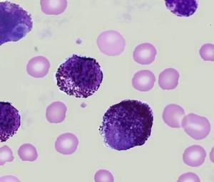
How do we determine if your pet has a mast cell tumor? Fortunately, mast cells are very distinct cells with deep purple granules in their cytoplasm. They are easily identified by a simple test called a fine needle aspirate. A needle is inserted into the lump in question and the plunger of the syringe is pulled back to obtain some cells. These cells are then squirted onto a glass slide, stained, and examined under the microscope. If mast cells are identified, surgical removal of the mass is the next step.
Mast cell tumors come in two forms: low grade and high grade. Tumors cannot be graded by the fine needle aspirate described above. The tumor and surrounding skin must be examined by an experienced pathologist (a doctor who examines biopsied tissues) to obtain a grade. Low grade tumors generally hold a good prognosis if completely removed. It is important to take a large amount of skin around the tumor to make sure all cancer cells are removed. This is called the surgical margin, and a wide margin makes regrowth of the tumor or spread to other areas of the body much less likely than a very narrow surgical margin. This is often challenging since many mast cell tumors seem to occur on the lower legs or face where wide margins are difficult to achieve. Drugs like steroids or antihistamines may be used to attempt to shrink the tumor before it is removed. High grade mast cell tumors are much more serious and indicate tumor spread into the lymph nodes or other organs such as liver or spleen.
Additional tests such as basic blood tests, abdominal ultrasound, aspiration of lymph nodes near the mass, and chest radiographs (x-rays) may also be recommended to screen or “stage” your pet’s mast cell tumor, especially if a high grade tumor is suspected or found. Chemotherapy options for treating mast cell tumors including prednisone, lomustine and vinblastine as well as a drug called Palladia are available as is radiation therapy. These treatments can extend lifespan and comfort for those pets who have multiple mast cell tumors or high grade or incompletely excised tumors.
Any time your pet develops a new lump, especially if the lump seems to change in size, please have your veterinarian perform an aspirate to determine the type of mass. If it is a mast cell tumor, timely removal with wide margins usually means a very good prognosis for most patients with low grade tumors. If your pet has previously had a mast cell tumor removed, be diligent about checking him for new tumors and let your vet know if any new masses are found so they can be evaluated. Mast cell tumors can be scary, but there are certainly treatments available if your pet should develop one of these tumors.
This blog brought to you by the Patton Veterinary Hospital serving Red Lion, York and the surrounding communities.
Location
Patton Veterinary Hospital
425 E Broadway
Red Lion, PA 17356
